Revision Knee Replacement
Revision knee replacement is performed to replace a failed or failing knee replacement.
Primary knnee replacement is generally a very successful operation. It can dramatically decrease pain, and increase quality of life. However some knee replacements can be painful, stiff, or unsatisfactory for many reasons.
Some may appear some time after the operation:
- Infection – infections can be very obvious, or can often be very subtle. Low-grade infections caused by bacteria such as Staph. epidermidis can cause pain, or progressive loosening of the components
- Loosening – the bond between the implants and the bone may fail, leading to pain and perhaps a sense of instability
- Wear – as the polyethylene bearing wears down, the knee can become increasingly unstable
These factors may be identified on serial X-rays performed during routine follow up. The surgeon who performed the original surgery should obtain yearly follow up x-rays to monitor the knee replacement.
However some factors may be related to the original operation:
- Implant malposition – subtle changes in implant position can make a drastic change to the function of a total knee replacement. Factors such as rotation of the components and the height of the prostheses can lead to poor movement of the tracking of the kneecap. This can lead to pain.
- Poor tissue balance – abnormal tightness or looseness of the ligaments due to incorrect bony cuts or incorrect surgical releases can lead to pain, stiffness, or a feeling of giving way.
A careful history and physical examination is very important. Following this, several investigations are performed, including x-rays, blood tests, and a CT scan. These are usually enough, but occasionally further tests may be needed including sampling the joint fluid for analysis, or testing the joint under anaesthetic. Frequently there may be more than one reason present, and it is important to identify them all.
Studies have shown that revision knee replacement is only successful when the causative problem has been properly identified. Revision surgery without a known cause is unlikely to be successful. Sometimes, multiple contributing factors exist.
In the case of infection, a staged procedure is preferred. This involves a first operation to remove the existing implants and cement, and clean the knee thoroughly. An antibiotic plastic spacer is then inserted to keep the joint under tension, and to deliver antibiotics to the local area. Intravenous antibiotics are then commenced for 6 weeks, and then ceased if the blood tests indicate the infection has cleared. The revision knee replacement is then inserted in a second operation.
Patients who are sufficiently troubled by their knee symptoms are considered for potential surgery. The gains of surgery must then be weighed up with the risks. Patients who are experiencing significant pain, and a reduction in quality of life, and have general health which is sufficiently robust, are suitable for surgery. Patients may require review by a physician or cardiologist, as well as an anaesthetist, for an opinion regarding fitness for surgery.
The problem knee without infection
The surgery is performed under spinal and/or general anaesthetic. Once the knee is prepared for surgery, the old incision is used to access the knee joint. Scar tissue build up is removed. The existing knee replacement is carefully removed, taking care to preserve as much residual bone as possible. Specimens are sent to confirm that no infection is present. The bony surfaces are thoroughly cleaned and any residual cement is removed. The new knee replacement is then positioned. This involved identifying the ideal position of the new prosthetic joint surfaces, and then substituting for any bone deficits using metal sections which join to the main prosthesis called augments. The femoral and tibial components are anchored to the bone with cement, and extensions which run into the bony canals called stems. The knee is the washed and closed.
The problem knee with infection
The surgery is performed under spinal and/or general anaesthetic. Once the knee is prepared for surgery, the old incision is used to access the knee joint. Scar tissue build up is removed. The existing knee replacement is carefully removed, taking care to preserve as much residual bone as possible. Specimens are sent to confirm the organism present. The bony surfaces are thoroughly cleaned and any residual cement is removed. An antibiotic spacer is made by combining high doses of antibiotic, and bone cement. This makes a plastic shape which mimics the shape of the knee joint, which leaches out antibiotic over several weeks, and greatly increases local antibiotic concentrations. Limited movement and weight bearing is possible with a spacer. The knee joint is then closed.
Intravenous antibiotics are then commenced for 6 weeks. Progress is measured via blood tests. Once these blood test return to normal, antibiotics are ceased. Repeat blood tests are performed after one to two weeks after cessation of antibiotics to demonstrate that the infection has definitively cleared. If these are normal, the second stage is planned.
At the second stage, the patient is again anaesthetised and prepared. The spacer is removed and the knee throughly cleaned. Specimens are again sent to confirm the absence of infection. The new knee replacement is then positioned. This involved identifying the ideal position of the new prosthetic joint surfaces, and then substituting for any bone deficits using metal sections which join to the main prosthesis called augments. The femoral and tibial components are anchored to the bone with cement, and extensions which run into the bony canals called stems. The knee is the washed and closed.
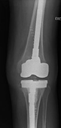
A revision total knee prosthesis.
Ideally, your original surgeon should monitor your progress carefully and identify any problems. They are well placed to treat any problems, and have good knowledge of your original surgery. However, some patients may benefit from a second opinion regarding their knee. Ensure that you seek an opinion from a surgeon specifically trained in revision surgery, and with adequate experience in performing this difficult surgery.
Generally, the risk involved with revision surgery are similar in nature to that of first-time knee replacement, but with higher magnitude. The risks of revision knee replacement include (but are not limited to):
- Infection – rare but potentially catastrophic. The infection rate in revision knee replacement surgery is around 2-4%. Infection may need further operations to wash out or revise the implants. In cases where the infection cannot be cleared despite removing the implants can end up requiring knee fusion or even amputation.
- Bleeding – blood transfusion may be required following surgery.
- Stiffness – persistent stiffness following revision surgery may occur. Usually, stiffness prior to surgery determines whether the knee will be stiff after revision. Most patients achieve more motion after revision surgery however.
- Pain – Occasionally patients are left with pain after the surgery as settled.
- Wear – the polyethylene in the knee can wear with time, requiring surgery. Sometimes a new polyethylene can be inserted, but occasionally the wear is associated with loosening of the metal components. In this situation, the knee replacement will have to be revised.
- Loosening – the bond between the metal and the bone can fail, leading to loosening and pain. This requires repeat revision surgery.
- Dislocation or instability – the components of the knee replacement may jump or slide abnormally, leading to giving way of the joint. This may require revision of the knee replacement to a design with more intrinsic stability.
- DVT/PE – Deep Vein Thrombosis (DVT) involves formation of blood clot in the deep calf veins. This may occur after surgery, trauma, or even spontaneously. Knee replacement is associated with a higher risk of DVT. Special precautions are usually taken, including compression socks, pneumatic pumps, and either blood thinning tablets or injections. Even despite these measures, DVT can occur. DVT by itself causes calf pain and swelling, but its most concerning consequence is when the clot breaks free and travels to the lungs (called Pulmonary Embolism or PE), causing shortness of breath or chest pain. Very occasionally this can be serious or even life-threatening.
- Fracture – a very rare complication. If this occurs, further surgery or splinting may be required.
- Injury to nearby nerves and blood vessels – very rare, but may be associated with impaired log term function.
- Anaesthetic problems – Anaesthetic agents have been associated with allergic and anaphylactic reactions. In addition, the medications can depress the function of the heart and lungs. In older or more prone patients this may lead to heart attack, stroke, or cardiac failure.
In well trained hands, revision knee replacement is a very successful operation. However, the risks are greater than those in primary knee replacement. There is an increased risk of stiffness, fracture, and infection.
The infection risk in the first total knee replacement is around 1-2%. In revision surgery this is around 3-4%. In surgery performed to clear infection in a total knee replacement, the success rate is around 80-90%.
The reason for revision also determines the success of the surgery. Patients who have surgery for unstable knees, those with worn plastic, or with loose implants generally do very well. Patients revised for stiffness due to excess scarring around the knee are less successful.


